Case Studies

Real-world evidence from the National PET Imaging Platform

The National PET Imaging Platform (NPIP) is enabling researchers and clinicians across the UK to push the boundaries of what’s possible with total-body PET imaging. From uncovering new diagnostic pathways to making previously off-limits scans safely available to patients, our case studies showcase the real impact this technology is having on research and patient care.

Browse the case studies below to see how total-body PET is being used across different disease areas, and get in touch if you’d like to explore how it could support your own research.
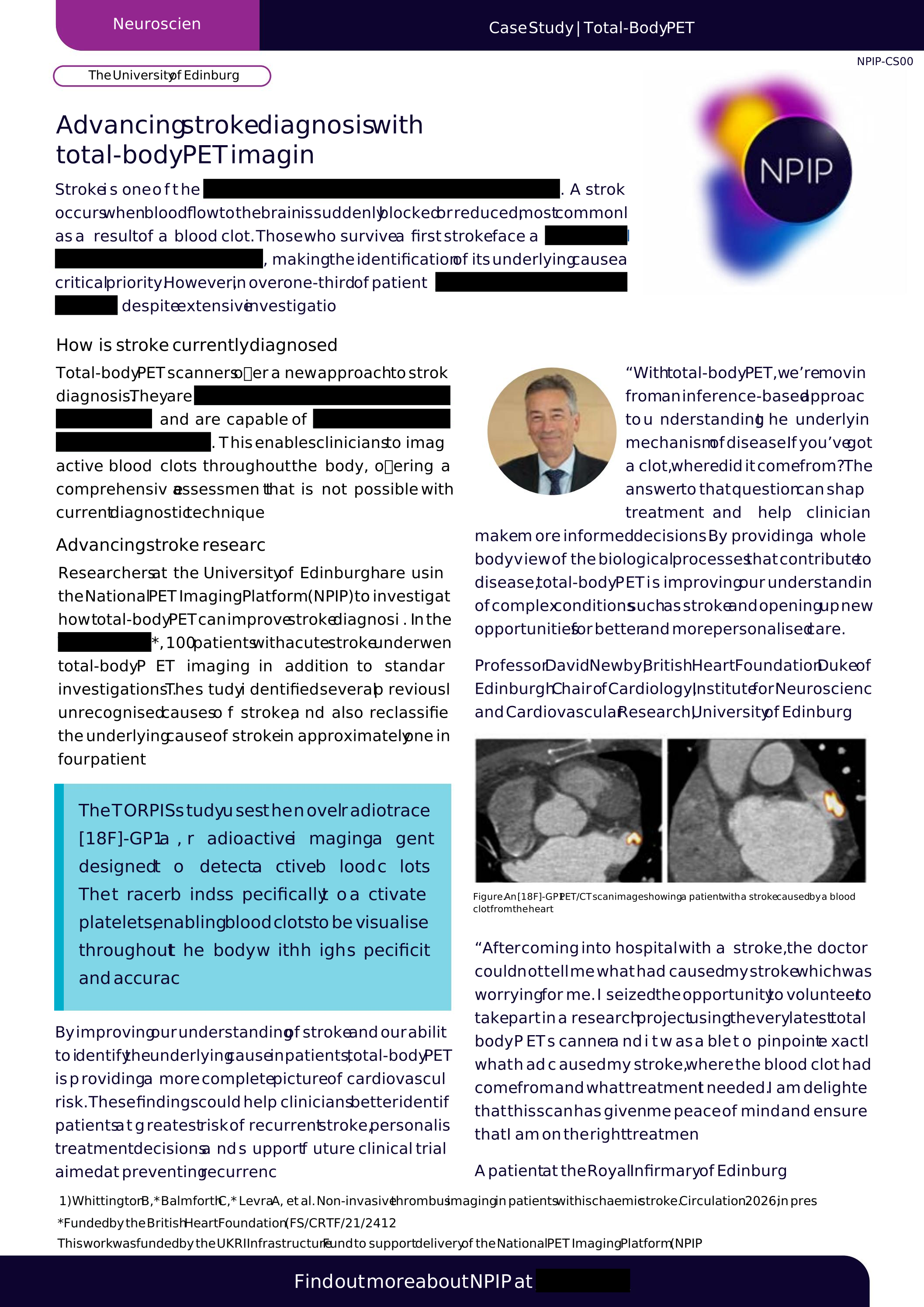
Endometriosis: Transforming endometriosis research with non-invasive total-body PET imaging

The University of Edinburgh

Endometriosis can take up to nine years to diagnose, and for many patients, surgery is currently the only definitive option. At the Royal Infirmary of Edinburgh, researchers are using total-body PET to trial two novel radiotracers, GP1 and FAPI, that target the distinct bleeding and scarring processes behind different types of endometriosis lesions. The study could pave the way for a faster, non-invasive diagnostic pathway, reducing unnecessary surgeries and improving understanding of how the condition develops and responds to treatment.

If you don’t see the case study below, click here.

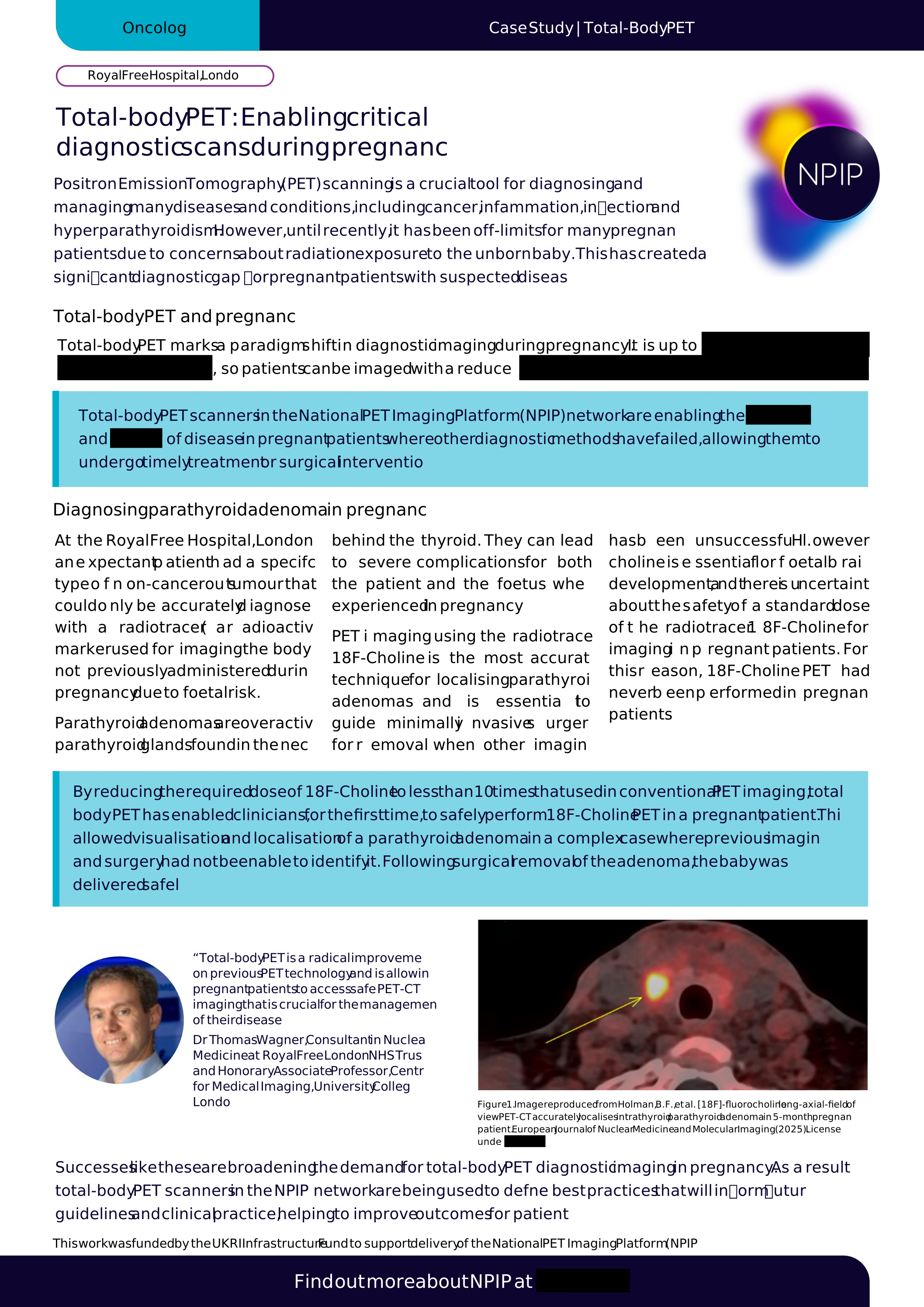
Oncology: Total-body PET enabling critical diagnostic scans during pregnancy

Royal Free Hospital, London

PET imaging has long been restricted in pregnancy due to concerns about radiation exposure to the unborn baby, creating a diagnostic gap for expectant patients with suspected disease. At the Royal Free Hospital, total-body PET’s greater sensitivity allowed clinicians to safely reduce the radiotracer dose and, for the first time, perform an 18F-Choline PET scan in a pregnant patient. This enabled the localisation of a parathyroid adenoma that previous imaging had failed to detect, guiding successful surgery and a safe delivery.

Case study will be available soon.

Total-body PET imaging is revolutionising medical imaging, providing a more complete picture of health for patients.

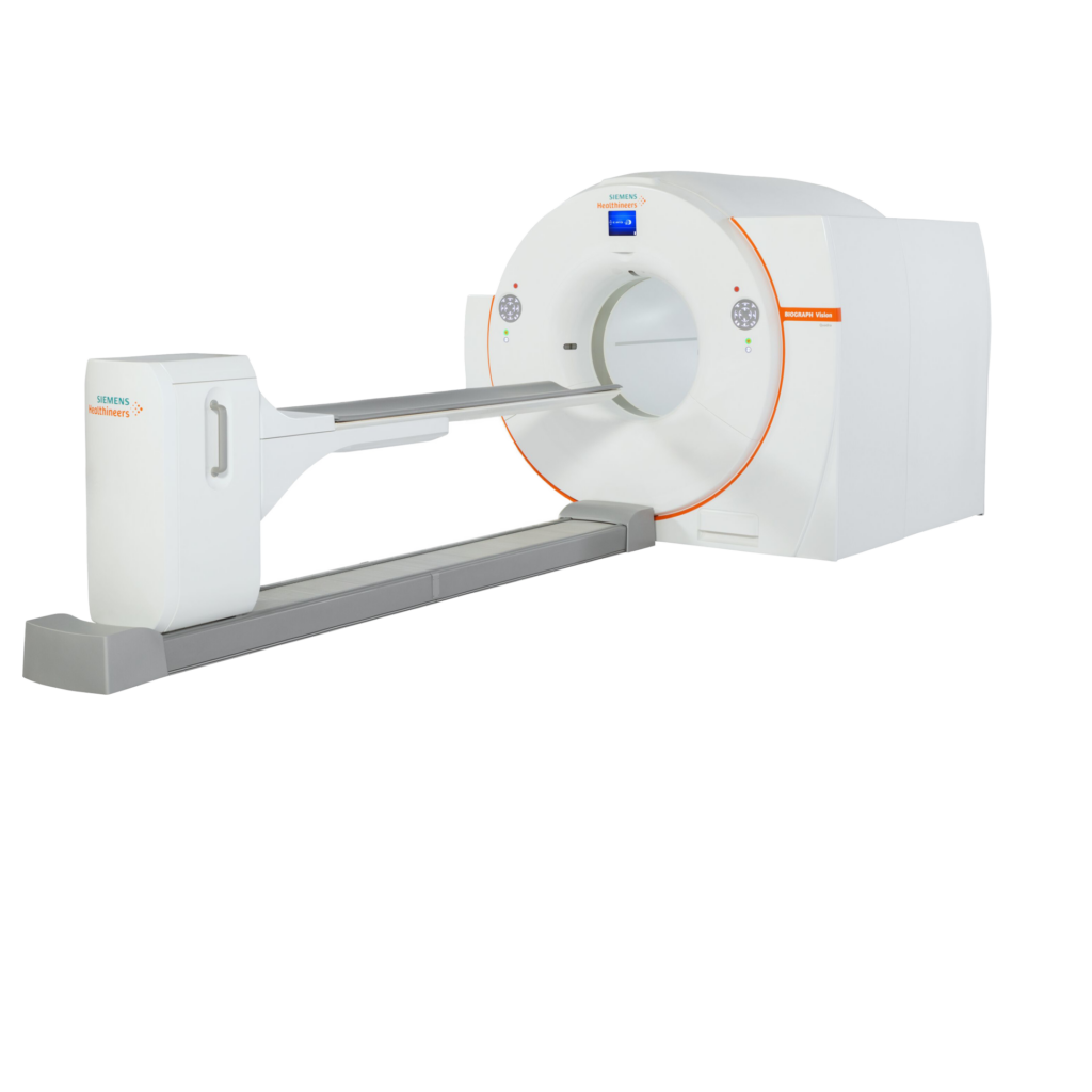
A total-body PET scan is a medical imaging technique that uses a small amount of radioactive material and a special camera to create detailed images of the inside of the body. A patient is injected with a small amount of radioactive tracer. The PET scanner then detects the gamma-ray emissions produced by the tracer as it accumulates in different parts of the body.

circle-shape

Our Partners

NPIP is created and delivered by internationally renowned players in medicine, science, and academia. Find out more about our partners.


Join the Platform


Interested in discussing these case studies or exploring how total-body PET could support your own research? Get in touch with the NPIP team — we’d love to hear from you.

Get in touch